Friday 2 October 2015

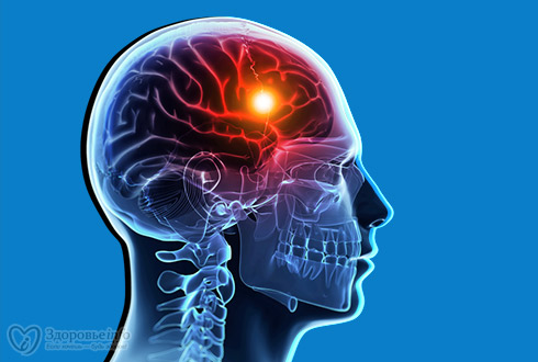
                   ANTI EPILEPTIC DRUGS


CLASSIFICATION


(ABCD SHIP)


A liphatic carboxylic acid
B  enzodiazepines
C  yclic GABA analogue
D  eoxy barbiturates

S   uccinimide
H  ydantoin
I   minostilbene
P  henyltriazene


Possible mechanism for seizure are:


          decreased inhibitory synaptic activity (i.e. GABA)                 
        (GABA antagonist triggers Sz)

      #  increased excitatory activity (i.e. Glutamate) 
            (Agonist triggers Sz)


Treatment

2 voltage-gate ion channels blocked for treatment of Sz


$ voltage-gated Na channels


  DRUGS BLOCKING Na CHANNEL

  (Mnemonic: Zodium ChannelsPartially Locked)

1. henytoin

2. arbamazepine 

3. amotrigine

4. onisamide 

 (Mnemonic: Possibly Voltage-gated Na Target)

5. P henobarbital

6. V alproate

7. T opiramate


$ T-type Ca channels


   DRUGS BLOCKING Ca CHANNEL
 its the receptor that governs oscillatory responses in thalamic neurons

1. Gabapentin


2. Pregabalin


drugs inhibiting oscillatory current b/w thalamus&cortex

(Mnemonic: Ethan sux because he's always absent from school and that's why Valerie won't datehim, but she oscillates on the decision)

3. Ethosuximide

4. Valproate

drugs enhancing GABA-ergic neurotransmission


post synaptically

  
     1.  B zd

     2.  B arbiturate

     3.  T opiramate

       
   # pre synaptically

     (Mnemonic: Tia is always gabbing in class so we block the door and leave her in the hallway/cleft) 

   1.Tiagabine


 Drug inhibiting degradation of GABA by GABA aminotransferase

  (Mnemonic:Viva GABA =ViGABAtrin leaves GABA in cleft)

  
    1.Vigabatrin


        DRUG  OF  CHOICE


TONIC CLONIC SEIZURE

1. Valproate

2. Carbamazepine

3. Phenytoin


ABSENCE SEIZURE


1. Ethosuximide

2. Valproate  

MYOCLONIC SEIZURE

1.Valproate


DRUG INDUCED Sz

1. Diazepam

2. Lorazepam

3. Phenobarbital


ADRs to be followed.. .. .. 

Stay cool!!
adieu..


Tuesday 5 May 2015

Stroke

Stroke is a term used to describe an abrupt onset of local neurological deficit that lasts at least 24 hours and is presumed to be of vascular origin.

CLASSIFICATION & ETIOLOGY
·        Ischemic stroke  <associated with local thrombus formation resulting in the occlusion of cerebral artery>

1.     Atherosclerotic cardiovascular disease
2.   Penetrating artery disease
3.   Cardiogenic embolism
4.   Cryptogenic stroke
5.   Other unusual causes
a)  Prothrombin state
b)  Arteritis
c)   Migraine
d)  Drug abuse

·       Hemorrhagic stroke <associated with uncontrolled B.P. and thrombolytic therapy>

1)    Intra parenchymal  (occurs when blood vessel ruptures within the brain parenchyma resulting in the formation of hematoma)
2)  Sub arachidonic  (occurs when blood enters into subarachidonic spaces due to trauma)

RISK FACTORS
·       Non modifiable risk factors or risk markers
1)    Age
2)  Gender
3)  Race
4)  Family history
5)  Low birth weight
·       Modifiable risk factors
1)    Hypertension
2)  Atrial fibrillation
3)  Diabetes
4)  Other cardiac disease
5)  Dyslipidemia
6)  Cigarrete smoking
7)  Alcohol
8)  Sickle cell disease
9)  Post-menopausal hormonal therapy
10)        Life style factors-obesity, sedentary lifestyle, diet

PATHOPHYSIOLOGY
ISCHEMIC STROKE

Accounts for 88% of strokes. Due to local thrombus formation or emboli that occlude a cerebral artery.
Cerebral ATH is a causative factor in most cases, while 30% is due to unknown etiology. An emboli can arise from intra or extra cranial arteries.
20% emboli arise from heart.
In Carotid ATH plaques may rupture causing collagen exposure, platelet aggregation and thrombus formation.
The cardiogenic embolism stasis of blood flow in the atria or ventricle
The final result of thrombus and emboli is atrial occlusion

HEMORRGHIC STROKE 

Sub arachidonic hemorrhage results from trauma or rupture or arteriovenous malformation (AVM)
Intra cerebral hemorrhage results from ruptured blood vessels within the brain parenchyma.

SIGNS & SYMPTOMS
·       Cognitive deficit
·       Patient won’t be able to give relevant history
·       One side weakness
·       Inability to speak
·       Loss of vision
·       Vertigo
DIAGNOSIS
·       CT Scan
·       MRI
·       Carotid Doppler studies
·       ECG to determine A.F.
·       Transthoracic echocardiogram
·       Trans esophageal echocardiogram


Thursday 30 April 2015

           Undescended testicle

Undescended testicle occurs when one or both testicles fail to move into the scrotum before birth.

Also called as Cryptorchidism; Empty scrotum - undescended testes; Scrotum - empty (undescended testes); Monorchism; Vanished testes - undescended; Retractile testes

Causes
Most of the time, a boy’s testicles descend by the time he is 9 months old. Undescended testicles are fairly common in infants who are born early. The problem occurs less often in full-term infants.
Some babies have a condition called retractile testes and the health care provider may not be able find the testicles. In this case, the testicle is normal but is pulled back out of the scrotum by a muscle reflex. This is able to occur because the testicles are still small before puberty. The testicles will descend normally at puberty and surgery is not needed.
Testicles that do not naturally descend into the scrotum are considered abnormal. An undescended testicle is more likely to develop cancer, even if it is brought into the scrotum with surgery. Cancer is also more likely in the other testicle.
Bringing the testicle into the scrotum can improve sperm production and increase the chances of good fertility. It also allows the health care provider to do an exam for the early detection of cancer.
In other cases, no testicle may be found, even during surgery. This may be due to a problem that occurred while the baby was still developing before birth.

Symptoms

Most of the time there are no symptoms other than the absence of the testicle in the scrotum. (This is called an empty scrotum.)

Exams and Tests

An exam by the health care provider confirms that one or both of the testicles are not in the scrotum.
The health care provider may or may not be able to feel the undescended testicle in the abdominal wall above the scrotum.
Imaging tests, such as an ultrasound or CT scan, may be done.


Possible Complications


·         Damage to the testicle from surgery Infertility later in life
·         Testicular cancer in both testes


Treatment


In most cases, the testicle will descend without treatment during the child’s first year. If this does not occur, treatment may include:
·         Hormone injections (B-HCG or testosterone) to try to bring the testicle into the scrotum
·         Surgery (orchiopexy) to bring the testicle into the scrotum. This is the main treatment.