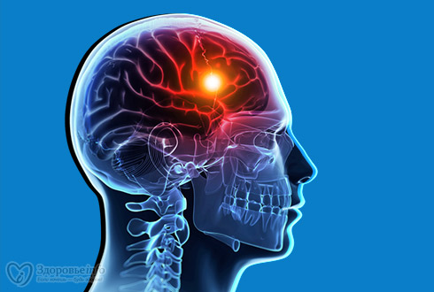
Stroke
 Stroke is a term used to describe an abrupt onset of local neurological
deficit that lasts at least 24 hours and is presumed to be of vascular origin.
Stroke is a term used to describe an abrupt onset of local neurological
deficit that lasts at least 24 hours and is presumed to be of vascular origin.
CLASSIFICATION
& ETIOLOGY
· Ischemic stroke <associated with local thrombus
formation resulting in the occlusion of cerebral artery>
1. Atherosclerotic
cardiovascular disease
2. Penetrating artery
disease
3. Cardiogenic
embolism
4. Cryptogenic stroke
5. Other unusual
causes
a) Prothrombin state
b) Arteritis
c) Migraine
d) Drug abuse
· Hemorrhagic stroke <associated with
uncontrolled B.P. and thrombolytic therapy>
1) Intra parenchymal (occurs when blood vessel ruptures within the
brain parenchyma resulting in the formation of hematoma)
2) Sub arachidonic (occurs when blood enters into subarachidonic
spaces due to trauma)
RISK
FACTORS
· Non modifiable risk factors or risk
markers
1) Age
2) Gender
3) Race
4) Family history
5) Low birth weight
· Modifiable risk factors
1) Hypertension
2) Atrial fibrillation
3) Diabetes
4) Other cardiac disease
5) Dyslipidemia
6) Cigarrete smoking
7) Alcohol
8) Sickle cell disease
9) Post-menopausal
hormonal therapy
10)
Life style factors-obesity, sedentary lifestyle, diet
PATHOPHYSIOLOGY
ISCHEMIC
STROKE
Accounts for 88% of strokes. Due to local thrombus formation or emboli
that occlude a cerebral artery.
Cerebral ATH is a causative
factor in most cases, while 30% is due to unknown etiology. An emboli can arise
from intra or extra cranial arteries.
20% emboli arise from heart.
In Carotid ATH plaques may
rupture causing collagen exposure, platelet aggregation and thrombus formation.
The cardiogenic embolism stasis
of blood flow in the atria or ventricle
The final result of thrombus and emboli is atrial occlusion
HEMORRGHIC
STROKE
Sub arachidonic hemorrhage results from trauma or rupture or
arteriovenous malformation (AVM)
Intra cerebral hemorrhage results from ruptured blood vessels within the
brain parenchyma.
SIGNS
& SYMPTOMS
 · Cognitive deficit
· Patient won’t be
able to give relevant history
· One side weakness
· Inability to speak
· Loss of vision
· Vertigo
DIAGNOSIS
· CT Scan
· MRI
· Carotid Doppler
studies
· ECG to determine
A.F.
· Transthoracic
echocardiogram
· Trans esophageal
echocardiogram